Case Report: An Interesting Case of Intracranial Tuberculoma with Spinal Tuberculous Spondylitis in a Patient with Muscular Dystrophy
Jasmine kalyani P*, Saravanan S , Sankar Narayanan, Ravi
Department of Neurology, Tirunelveli Medical College Palayamkottai, Tirunelveli, Tamil Nadu, India
- *Corresponding Author:
- Jasmine Kalyani
Department of Neurology, Tirunelveli Medical College Palayamkottai, Tirunelveli, Tamil Nadu, India
Tel no: 9442370976
E-mail: jasminekalyani31@gmail.com
Received Date: December 03,2021; Accepted Date: December 16,2021; Published Date: December 23,2021
Citation: Jasmine k aly ani P , Sar a v anan S, Nar a y anan S, Ra vi (2021) Case R eportI:n tAenre sting Case of Intracranial Tuberculoma with Spinal
Tuberculous Spondylitis in a Patient with Muscular Dystrophy. J Neurol Neurosci Vol:12 No:9
Abstract
This case describes an unusual or novel occurrence. An unexpected association between intracranial tuberculoma and spinal tuberculosis. This is an unexpected event in the course observing and treating a patient with muscular dystrophy [1]. We report a case of combination of spinal tuberculous spondylitis [2] and intracranial tuberculoma [3] which is extremely rare, so far; only five cases have been reported in the literature. 32-year-old female who is a known case of muscular dystrophy presenting with fever, diplopia, low back ache and motor weakness of both upper and lower extremities. Based on magnetic resonance imaging and polymerase chain reaction, we diagnosed as tuberculoma. She was started on conventional antituberculous [4] treatment and steroids [5].
Keywords
Intracranial tuberculoma; Brain; Spine; Weakness and Magnetic resonance imaging
Highlights
The association of potts spine with intracranial tuberculomas is extremely rare. Up to date, there are only few cases which have been reported in the literature [6,7]. Intracranial tuberculomas associated with intramedullary tuberculomas only about 5 cases has been reported worldwide.
Introduction
Tuberculosis which is a catastrophic infectious disease caused by Mycobacterium tuberculosis presents with extra pulmonary forms with drug resistance in the recent days. Most common manifestations of tuberculosis in the CNS [8] are tuberculous meningitis [9] and intracranial tuberculoma. Brain is far more commonly affected than the spinal cord. Infratentorial tuberculomas [10] are more frequent in children, whereas lesions are mostly supratentorial [11] in adults. Tuberculous spondylitis, also known asPott disease, refers to vertebral body osteomyelitis and intervertebral discitis fromtuberculosis (TB). The spine is the most frequent location ofmusculoskeletal tuberculosis almost 50%. In spinal tuberculosis, characteristically, there is destruction of the intervertebral disk space and the adjacent vertebral bodies, collapse of the spinal elements, and anterior wedging leading to the characteristic angulation and gibbus. Discitis [12] and/or osteomyelitis usually affect the lower thoracic and upper lumbar levels of the spine.
Spine is involved due to hematogenous spread [13] that can occur via both arteries and veins, resulting in different patterns of infection.
Spread [14] through the anterior arterial arcade that richly supplies the subchondral paradiscal bone results in infection anterosuperiorly and anteroinferior, adjacent to the disc. In adults and particularly older individuals the disc is conspicuously spared due to its sparse vascularity. In contrast, in younger individuals, especially children, the disc may be involved early as it has a far richer blood supply.
Infection then spreads beneath the longitudinal ligaments and can lead to infection of adjacent vertebral bodies.
Gradual anterior collapse typically results in an acute kyphotic or gibbus deformity [15]this angulation, coupled with epidural granulation tissue [16] and bony fragments, can lead to cord compression [17].
Central involvement: Spread via thevenous plexus of Batson [18]typically results in infection arising centrally within the vertebral body. This is more common in older individuals.
Gradual collapse can result in vertebra plana and acute kyphotic orgibbus deformity. This angulation, coupled with epidural granulation tissue and bony fragments, can lead to cord compression.
Posterior involvement, also known as an appendiceal [19] is also due to venous hematogenous spread via the posterior venous plexuspattern. : Synovial involvement [20] is relatively rare but can be seen involving facet joints and atlantoaxial and atlanto-occipital joints. In late-stage spinal tuberculosis, large paraspinal abscesses can develop without severe pain or frank pus or prominent inflammatory signs and symptoms; thus "cold abscess" [21].
Incidence of neurologic complications in spinal tuberculosis varies from 10 to 43 %
Clinical presentation
A 32-year-old female immunocompetent patient who is a known case of muscular dystrophy came with signs of headache, diplopia, and weakness in both upper and lower limbs. She had severe lowback ache and progressive weakness of both lower limbs. There was also the history of the evening rise of temperature, loss of weight and appetite. On examination, she had quadriparesis with a power 2/5 proximally and 4/5 distally in upper limbs and 2/5 in the lower limbs both proximally and distally.
Both superficial and deep tendon reflexes were absent and plantar was extensor. Tone was hypotonia in both lower limbs. Generalised wasting was present. She presented with sensory involvement in the form of reduced perception of all modalities from T 10 segmental level. There was excruciating radiating pain from the lower back down to both legs which was aggravated by movement, coughing and straining at stools. Regarding higher mental function she was conscious, oriented, memory, speech and behaviour were normal. She had right lateral rectus palsy.
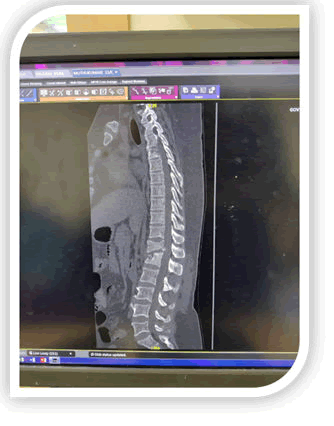
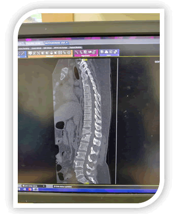
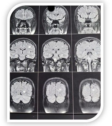
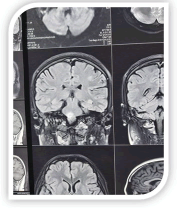
On the evaluation, Erythrocyte Sedimentation Rate (ESR) was 40 mm/1st h, chest X-ray was normal. Sputum examination for Acid Fast-Bacilli (AFB) could not be done as a patient had no cough. Mantoux test was positive and Cerebrospinal Fluid (CSF) was clear, with 4 white blood cell/dL (100% lymphocytes), sugar 52 mg/dL, proteins 37 mg/dL, chloride 89 mmol/L. AFB staining was negative in CSF with adenosine deaminase activity levels of 7 IU/L. Magnetic Resonance Imaging (MRI) brain showed well-defined ring-enhancing lesions with perilesional edema in pons, suggestive of tuberculoma [Figures 1 and2]. X-Ray spine showed bony destruction of D12 and anterior wedge fracture of L1. Spine CT showedspondylotic destruction of D12 to L1 vertebral bodies, compression of conus medullaris and Rt sacroiliitis.
Discussion
Central nervous system tuberculosis is rare, affecting 0. 5-2% of patients with systemic tuberculosis.
Tuberculoma is a peculiar manifestation of tuberculosis that might occur in any solid organ of the body. It is, usually, formed by conglomeration of several miliary tubercles [22], with the centre of the conglomeration becoming caseous. Caseous material gets inspissated and sometimes liquefied. A thick capsule may form around these lesions.Concurrent occurrence of intracranial tuberculomas along with spinal tuberculosis is extremely rare.
The case presented here was diagnosed in the background ofmuscular dystrophy. The chest X-ray of this patient was normal. The patient had elevated ESR and was symptomatic in the form of loss of weight and appetite with occasional evening rise of temperature.
Magnetic resonance imaging is the optimal measure because it shows location, size, and number of lesions and the presence of degeneration and necrosis.
The differential diagnosis of tuberculomas includes granulomas such as cysticercal granulomas and neoplastic lesions such as astrocytoma, metastasis or lymphoma. In this case, the clinical presentation and size of the lesion combined with the classical ring enhancement and surrounding edema was thought to be typical of a tuberculous granuloma. Associatedpotts spine also confirms the aetiology. Paraplegia is the devastating complication found in this patient, Hodgson, in his classical paper on Pott's paraplegia, classified paraplegia into two groups according to the activity of the tuberculous infection. These two groups were paraplegia of active disease (early-onset paraplegia) and paraplegia of healed disease (late-onset paraplegia). Early-onset paraplegia develops in the active stage of spinal tuberculosis and requires active treatment. This type of paraplegia has a better prognosis and is frequently seen in adults with Pott's spine Late-onset paraplegia may develop two to three decades after active infection. It is often associated with marked spinal deformities.
Conclusion
We report a case of concurrent occurrence of spinal tuberculous spondylitis and intracranial tuberculomas in a patient with muscular dystrophy. This case is being presented because of extreme rarity. Medical therapy is generally advocated as the initial treatment. High index of suspicion is needed to diagnose extrapulmonary tuberculosis particularly in a developing country like India where the prevalence of all forms of tuberculosis is very high 5. 05 /1000 before considering other etiologies like demyelination.
References
- Muscular dystrophies: Prof Eugenio Mercuri, PhD Prof Carsten G Bönnemann, PhD, Prof Francesco Muntoni, MD Published:November 30:19
- Michael P Hartung Tuberculous spondylitisLast revised ? on 04 Sep 2021 Ravindra
- Kumar Garg R. Tuberculosis of the central nervous system
- Development of antituberculous drugs: current status and future prospects[Article in Japanese]Haruaki Tomioka , Kenji NambaAffiliations expandPMID: 17240921
- Harry Shubin M D, Robert E, Lambert M D, Charles A, Heiken M D et al. (1959) Steroid therapy and tuberculosis.JAMA170:1885-1890
- Keklikoglu H, Yildiz’s C, Yoldas T, and Güler S (2008) “Central nervous system tuberculoma and pott’s disease: case report ”J Neurol Sci 25:142–147
- Tuberculosis of Central Nervous System Manish Modi, Abhishek GargPublished:08 September 2017
- Tuberculous meningitis Robert J. Wilkinson, Ursula Rohlwink, Usha Kant Misra, Reinout van Crevel, Nguyen Thi Hoang Mai, Kelly E. Dooley, Maxine Caws, Anthony Figaji, Rada Savic, Regan Solomons & Guy E. Thwaites on behalf of the Tuberculous Meningitis International Research Consortium
- Tuberculoma: Tuberculomas are conglomerate caseous foci within the substance of the brain that develop from deep-seated tubercles acquired during a recent or remote period of bacillemia. From: Aminoff's Neurology and General Medicine (Fifth Edition), 2014
- Winarno ANS. Multiple intracranial tuberculomas: a rare case of cns tb
- Rajasekaran S, PhD, FRCS, MCh, Dilip Chand Raja Soundararajan, MS, Ajoy Prasad Shetty, MS, (2018) Spinal Tuberculosis: Current Concepts 5. First Published December 13
- Rebecca A, Clark, Sally L, Blakley, Greer D, Margaret H, et al. Hematogenous Dissemination of Mycobacterium tuberculosis in Patients with AIDS
- Burrill J, Christopher J, Bain G, Conder G, Andrew L, et al. (2007) Tuberculosis: A Radiologic Review Sep 1
- Rajasekaran S, Shanmugasundaram T K (1987) Prediction of the angle of gibbus deformity in tuberculosis of the spine. J Bone Joint Surg 69:503-509
- Sobti S, Grover A, Paul B, John S, Sarvpreet Singh Grewal1, Uttam B George2 Prospective randomized comparative study to evaluate epidural fibrosis and surgical outcome in patients undergoing lumbar laminectomy with epidural autologous free fat graft or gelfoam: A preliminary study
- Kumar GargR and Somvanshi DS. Spinal tuberculosis: A review the journal of spinal cord medicine
- Ping Zhen MD, PhD, Xu-sheng Li MD, Hao Lu MD, Single Vertebra Tuberculosis Presenting with Solitary Localized Osteolytic Lesion in Young Adult Lumbar Spines
- Sharath Chandra B J, Girish T U, Thrishuli P B. Primary Tuberculosis of the Appendix: A Rare Cause of a Common Disease
- NobukazuP,KenichiF,GembaAtsushiYaoShinjiOzakiKatsuichiroOnoSaeWadaYasuhiroFujiiTakumiKishimotoYoshifumiNambTuberculosis diagnosed by PCR analysis of synovial fluid
- Patel R, Gannamani V, Shay E, Alcid D. Spinal Tuberculosis and Cold Abscess without Known Primary Disease: Case Report and Review of the Literature
- Surendra K, Mohanb SA, Sharmac A. Journal of clinical tuberculosis and other mycobacterial diseasesMiliary tuberculosis: A new look at an oldfoepanel

Open Access Journals
- Aquaculture & Veterinary Science
- Chemistry & Chemical Sciences
- Clinical Sciences
- Engineering
- General Science
- Genetics & Molecular Biology
- Health Care & Nursing
- Immunology & Microbiology
- Materials Science
- Mathematics & Physics
- Medical Sciences
- Neurology & Psychiatry
- Oncology & Cancer Science
- Pharmaceutical Sciences


